During the division of a cell DNA replication and cell growth also take place. Living things may appear very different from one another on the outside but their cells are very similar.

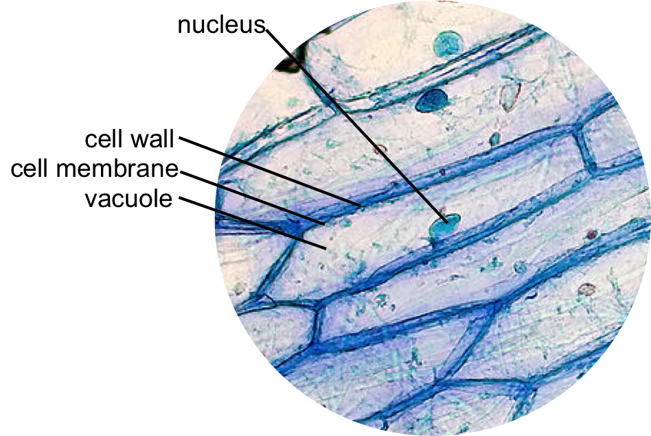
Epidermal Onion Cells Under A Microscope Plant Cells Appear Polygonal From The Cell Diagram Plant Cell Diagram Plant Cell
Stomata play an important role in gaseous exchange and photosynthesis.

. Making up all living material the cell is considered to be the building block of life. Compare the human cells on the left in Figure below and onion cells on the right in Figure below. Much later in 1831 Robert Brown an Englishman observed that all cells had a centrally positioned body which he termed.
The nucleus at the central part of the cheek cell contains DNA. When a drop of methylene blue is introduced the nucleus is stained which makes it stand out and be clearly seen under the microscope. They control by transpiration rate by opening and closing.
All these processes ie cell division DNA replication and cell growth hence. Determining time spent in different phases of the cell cycle The life cycle of the cell is typically divided into 5 major phases. The phases are listed below along with the major events that occur during each phase.
Viewed under a light microscope the nucleus appears only as a darker region of the cell but as we increase magnification we find that the nucleus is densely filled with a stew. In some of the plants stomata are present on stems and other parts of plants. When you are ready challenge your knowledge in the testing section to see what you have learned.
The nucleus a component of most eukaryotic cells was identified as the hub of cellular activity 150 years ago. Although the entire cell appears light blue in color the nucleus at the central part of the cell is much darker which allows it to be identified. Cryofixation and cryofracture techniques enabled us to describe the ultrastructure of these centrosome regions which are devoid of classical centriolar structures.
Aims of the experiment. Cycles of growth and division allow a single cell to form a structure consisting of millions of cells. Dinoflagellate centrosomes have been identified previously by using anti-β-tubulin antibody and CTR 210 an antibody raised against human cell centrosomes.
Stomata are the tiny openings present on the epidermis of leaves. Mount a Slide Look at Your Cheek Cells Lesson 3. 2 of 10 STEP 1 - Carefully cut an onion in half or.
In 1672 Leeuwenhoek observed bacteria sperms and red blood corpuscles all of which were cells. Lesson Description BioNetworks Virtual Microscope is the first fully interactive 3D scope - its a great practice tool to prepare you for working in a science lab. As a matter of fact observing onion cells through a microscope lens is a staple part of most introductory classes in cell biology - so dont be surprised if your laboratory reeks of onions during the first week of the semester.
A cell is the basic unit of the structure and function of living things. All forms of life are built of at least one cell. 101 CELL CYCLE Cell division is a very important process in all living organisms.
We can see stomata under the light microscope. We also describe. Once slides have been prepared they can be examined under a microscope.
To make observations and draw scale. This work is licensed under a Creative Commons Attribution-NonCommercial-NoDerivs 25 LicenseCreative Commons Attribution-NonCommercial-NoDerivs 25 License. The Biology Project Cell Biology Intro to Onion Root Tips Activity Activity Online Onion Root Tips.
One of the easiest simplest and also fun ways to learn about microscopy is to look at onion cells under a microscope. Explore topics on usage care terminology and then interact with a fully functional virtual microscope. It is easier to see nuclei under a light microscope with staining such as methylene blue.
To use a light microscope to examine animal or plant cells. 1665 observed a piece of cork under the microscope and found it to be made of small compartments which he called cells Latin cell small room. There are two microscope lesson activities in this blog for you to see the nuclei in animal cells and plant cells.
An onion a slide and cover slip a cotton bud some food colouring a plate to put the cotton bud on and of course a microscope. How are they similar.

Onion Cells Under A Microscope Requirements Preparation Observation Plant And Animal Cells Animal Cell Plant Cell

Onion Cells Google Images Ilustracoes Felipao

Onion Cells Cool Science Experiments Macro And Micro Microscopic Photography

Epidermal Onion Cells Under A Microscope Plant Cells Appear Polygonal From The Cell Diagram Plant Cell Diagram Plant Cell
0 Comments